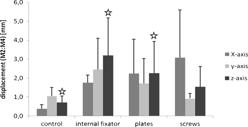
Fig. 3.
Displacement in millimetres (mm) of marker points two and four measuring the movement in the ruptured sacroiliac joint. Data shown are medium values from six different specimens after treatment with three loading cycles each. The three osteosyntheses are compared to the control pelvic ring without defects, p < 0.05

