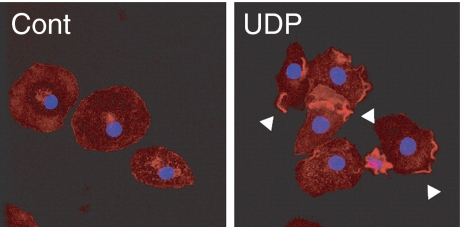
Fig. 1.
UDP induced local actin polymerization in microglia. Microglial cells were stimulated with or without UDP (100 μM) for 3 min. The cells were then fixed and stained for F-actin with Texas-Red phalloidin (red) and nuclei with DAPI (blue). In contrast to control cells (Cont), UDP induced F-actin aggregated in local cellular membranes, as indicated by arrowheads (UDP)

