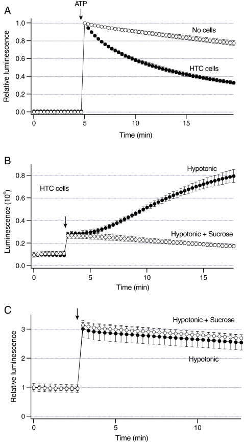
Fig. 1.
Increases in liver cell volume stimulate sustained ATP release.a Luminescence was measured from dishes with HTC cells (closed circles, n = 6 dishes) and no cells (open circles, n = 3 dishes). 500 nM ATP was added at arrow. Note that exogenous ATP is rapidly hydrolyzed in the presence of cells. b Luminescence was measured from dishes with HTC cells. Under basal conditions, these cells exhibit constitutive ATP release as indicated by non-zero luminescence values. Exposure to hypotonic solution (30% dilution) at arrow rapidly increased luminescence to a steady-state level (closed circles, n = 5 dishes). After ~2 min, luminescence gradually increased at a constant rate. In some experiments, cells were exposed to a solution that was a mixture of hypotonic solution and sucrose. This solution has the same osmolarity as control solution, and does not increase cell volume. Exposure to hypotonic + sucrose rapidly increased luminescence, but failed to further increase luminescence (open circles, n = 4 dishes). c Luminescence was measured from the dishes with no cells containing 20 nM ATP. Exposure to hypotonic solution at arrow rapidly increased relative luminescence (closed circles, n = 4 dishes). Similar changes were obtained after exposure to hypotonic + sucrose (open circles, n = 3 dishes)

