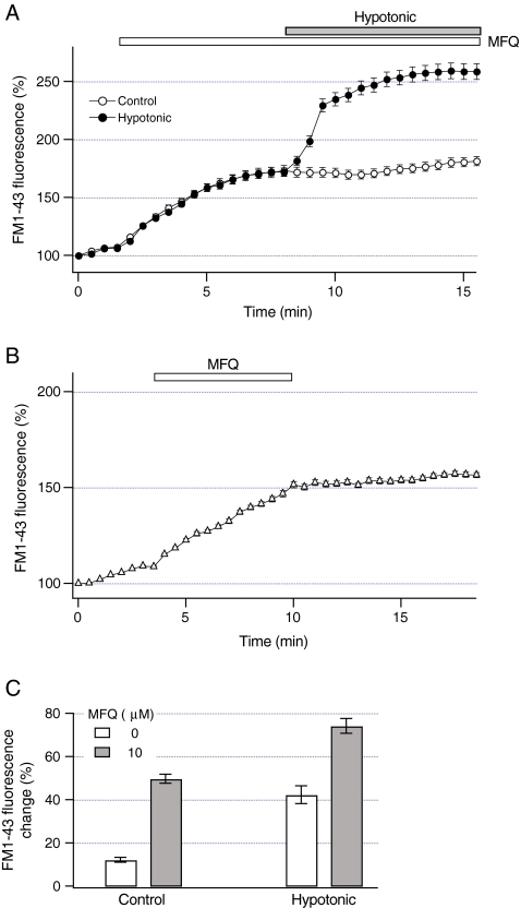
Fig. 6.
Mefloquine does not inhibit vesicular exocytosis. a FM1-43 fluorescence was measured in HTC cells after the exposure to 10 microM mefloquine (open circles, n = 8 cells). FM1-43 fluorescence was also measured from cells exposed to 10 microM mefloquine for 5 min before the exposure to hypotonic solution (30% dilution, closed circles, n = 13 cells). Note that mefloquine did not inhibit an increase in FM1-43 fluorescence evoked by hypotonic solution. b FM1-43 fluorescence was measured after the exposure to mefloquine (10 microM for 7 min, open bar). Note that removal of mefloquine did not decrease FM1-43 fluorescence (n = 13 cells). c Magnitude of constitutive exocytosis under control conditions was measured as a change in FM1-43 fluorescence that occurred 5 min after the exposure to 10 microM mefloquine (closed bar) or in the absence of mefloquine (open bar). The magnitude of exocytosis evoked by hypotonic solution was measured as a change in FM1-43 fluorescence that occurred 5 min after the exposure to hypotonic solution in the presence of 10 microM mefloquine (closed bar) or absence of mefloquine (open bar). Note that mefloquine did not inhibit exocytosis evoked by hypotonic solution. The number of analyzed cells ranged from 12 to 34

