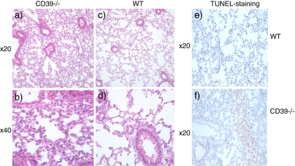
Fig. 3.
H&E staining of lung tissue collected after 45 min of full hepatic ischemia and 4 h of reperfusion. It is clearly visible that the Cd39-null animals (a, b) show increased alveolar edema compared with the wildtype controls (c, d). TUNEL-staining indicative of apoptosis demonstrates areas of apoptosis in the Cd39-null (f) lung tissue after 45 min of full hepatic ischemia and 4 h of reperfusion, when compared to the wildtype control (e)

