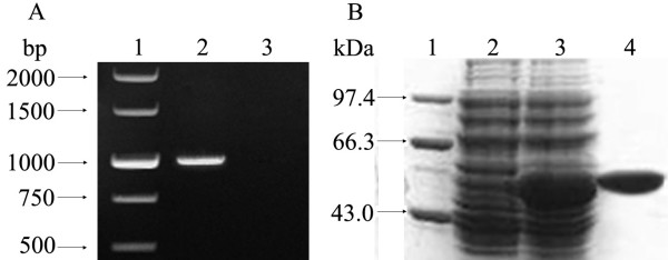
Figure 1.
Amplification and expression of the fliY gene and purification of the rFliY protein. Panel A, showing PCR analysis. Lane 1: DNA marker (TaKaRa, China); lane 2: the amplification segment of the entire fliY gene; lane 3: blank control. Panel B, showing SDS-PAGE analysis. Lane 1: protein marker (TaKaRa); lane 2: pET32a with no insertion of the fliY gene; lane 3: the expressed recombinant protein, rFliY; lane 4: the purified rFliY protein.

