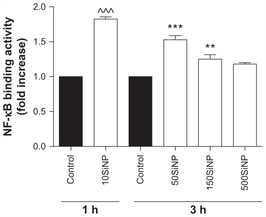
Figure 4.
Amorphous SiNP induce NF-κB DNA binding activity in primary human umbilical vein endothelial cells. Exposure of cells to 10 μg/mL 10SiNP for 1 hour induced a significant increase of the NF-κB activity when compared with the control. Also, exposure of 50 μg/mL 50SiNP and 150SiNP, but not 500SiNP, for 3 hours induced increased transcription factor activity. The NF-κB DNA binding activity induced by each nanoparticle treatment is expressed relative to the control sample.
Notes: All values are presented as the mean ± standard error of the mean of n = 4. By two-tailed Student’s t-test, ^^^P < 0.001 compared with the control. By one-way analysis of variance, Tukey–Kramer multiple comparison test: **P < 0.01 and ***P < 0.001 compared with the control. Filled bars correspond to cells which were not exposed to nanoparticles (control) and open bars to cells exposed to nanoparticles.
Abbreviation: SiNP, silica nanoparticles.

