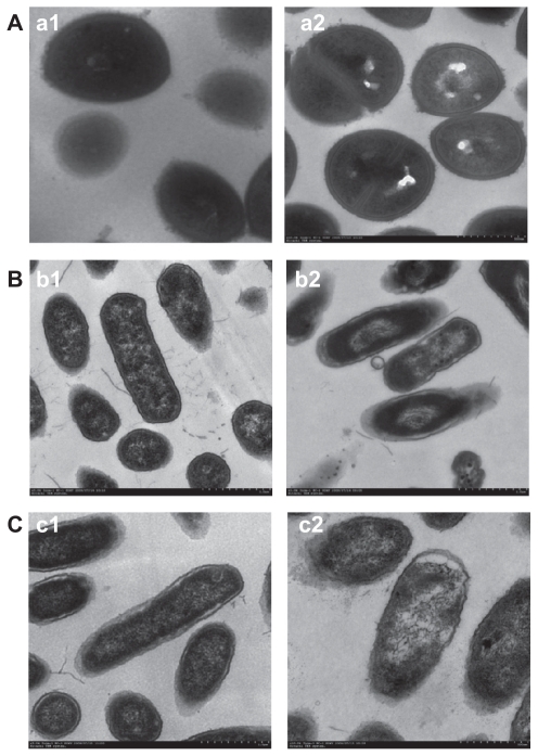
Figure 2.
Morphology and structure of bacterial cells under transmission electron microscopy. (A) a1, normal Staphylococcus aureus cells; a2, S. aureus cells treated by S-T-Gel. (B) b1, normal Escherichia coli cells; b2, E. coli cells treated by S-T-Gel. (C) c1, normal Pseudomonas aeruginosa cells; c2, P. aeruginosa cells treated by S-T-Gel.
Abbreviation: S-T-Gel, silver nanoparticles incorporated into thermosensitive gel.

