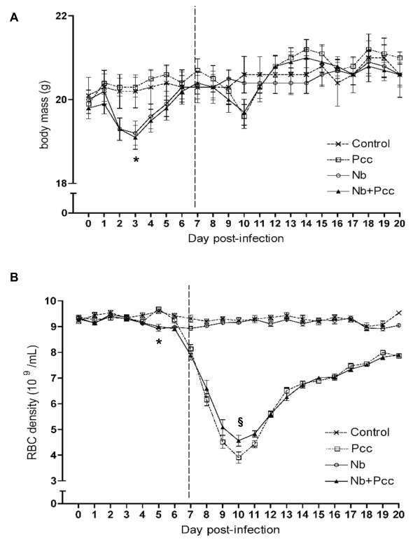
Figure 1.
Pathology, measured as loss of body mass (A) and red blood cell density (B). Mice were infected with 200 Nb L3 larvae and/or 105 Pcc-infected RBCs on day 0, then weighed daily to the nearest 0.1 g. Daily samples of blood were taken for flow cytometric analysis of RBC density. (A) Nb infection induced significant loss of body mass during the first week of infection (lower minimum body mass than mice without Nb; P < 0.0001). (B) Nb also induced significant loss of RBCs that week (lower minimum RBC density to 7 days pi than mice without Nb; P = 0.0037), whereas Pcc-induced RBC loss was mainly apparent during the second week of infection (lower minimum RBC density to 20 days pi than mice without Pcc; P < 0.0001). Nb co-infection significantly ameliorated this (higher minimum RBC density in co-infected compared to Pcc-infected mice; P = 0.0003). Means ± SEM from combined results of 3 independent experiments (each with 4-9 mice per group per time point) are shown. The vertical dashed line indicates the two time periods for which minimum weight and RBC density were analysed: up to 7 days pi, and 7-20 days pi, as described in Methods. The symbol * indicates statistical significance of the Nb main effect, and § indicates significance of the difference between co-infected and Pcc mice.

