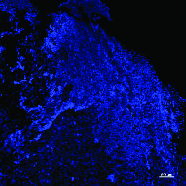
Figure 2.
Representative image from the middle ear mucosal biopsy of a child having a cochlear implant who has no history of middle ear disease. Child 21. FISH -EUB338 (Yellow), S. pneumoniae (green), H. influenzae (pink) and Hoechst 33342 (nuclei stain - blue). This maximum intensity projection (Z = 20.5 μm) demonstrates the normal mucosal tissue with no evidence of bacteria. Scale bar = 50 μm.

