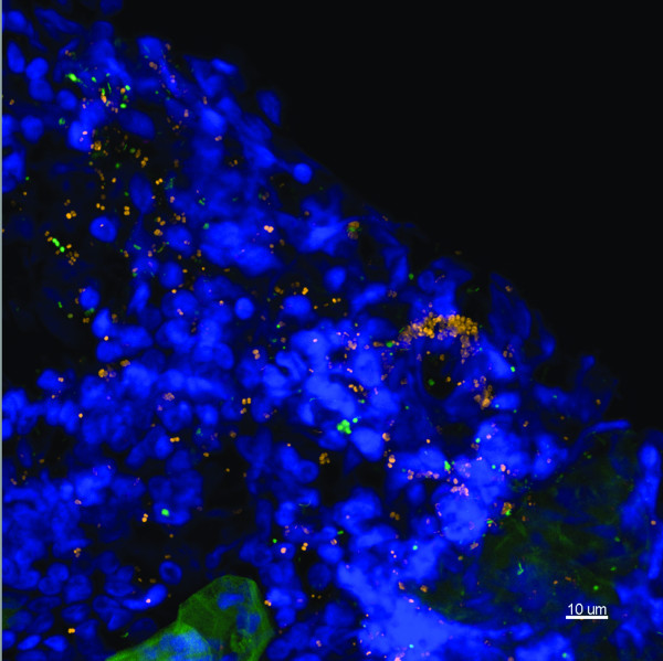
Figure 4.
Representative image of a mucosal biopsy from a child suffering from rAOM and COME. MEE was not present. Child 14. FISH probes included EUB338 (yellow), S. pneumoniae (green), negative for H. influenzae (pink), Hoechst 33342 (nuclei stain - blue). This is a maximum intensity projection (Z = 39 μm) showing multispecies biofilm covering the mucosa. The biofilm is seen to consist of S. pneumoniae and other unidentified bacteria. These bacteria are also interspersed within the tissue. Scale bar = 10 μm.

