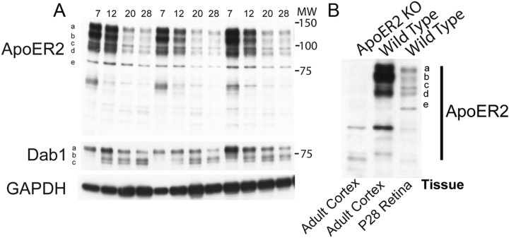
Figure 1.
Expression of ApoER2 in the developing retina. A, Retinas isolated at P7–P28 were subjected to Western blot analysis, revealing dramatic reductions in ApoER2 expression from P7 through P20. ApoER2 was found to exist in at least five forms (a–e), which all changed similarly across development. Molecular weight (MW, in kilodaltons) is indicated alongside the blots. GADPH levels revealed little difference in loading. B, Antibody specificity was determined by probing cortical lysates derived from adult ApoER2 KO and wild-type mice and comparing them to wild-type retinal lysate from P28. ApoER2 isoforms found in the adult cortex were identical to those found in the developing retina and were not found in ApoER2 KOs.

