Figure 4.

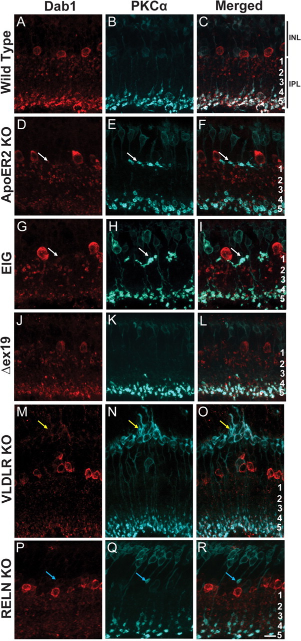
ApoER2 is important for rod bipolar and A-II amacrine morphogenesis. A-II amacrine and rod bipolar morphology was determined using Dab1 (red) and PKCα (blue) colabeling, respectively. A–C, Wild-type retinas had a typical distribution of ON rod bipolar axons in s5 and cell bodies present in the outermost portion of the INL. A-II amacrine dendrites were predominantly localized to s1–s2 and s5. D–F, A subpopulation of rod bipolar axons failed to penetrate the IPL in ApoER2 KO mice and formed multiple terminals along the border of the IPL and INL. Ectopic terminals were often associated with A-II amacrine dendrites (white arrows), which were also notably less abundant in s5. G–I, Similarly, ApoER2-EIG mice had rod bipolar axons that failed to penetrate the IPL and had reduced A-II amacrine dendrites in s5. J–L, ApoER2Δex19 mice had normal rod bipolar and A-II amacrine morphology. M–O, Although rod bipolar axons terminated properly in VLDLR KOs, structural remodeling of their dendrites occurred around sites of neovascularization (yellow arrows). A-II amacrine dendrites were also less distinct in the IPL, and remodeling occurred around sites of neovascularization. P–R, Failure of rod bipolar axons to penetrate the IPL is evident in Reln mutant mice (blue arrows), but axons did not form multiple terminals. Similar to VLDLR KOs, A-II amacrine dendrites throughout the IPL were less distinct. Scale bar: R (for A–R), 10 μm.
