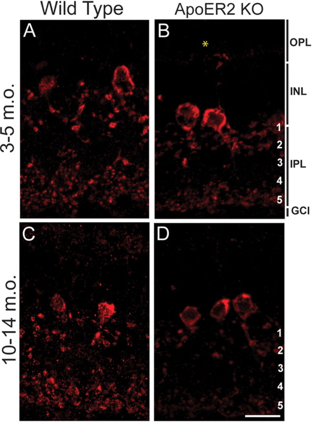
Figure 6.

Loss of ApoER2 is associated with altered A-II amacrine morphology in the aging retina. Dab1 (red) was labeled to visualize changes in A-II amacrine morphology in 3- to 5-month-old (m.o.) and 10- to 14-month-old wild-type and ApoER2 KO mice. A, C, Typical bistratified A-II amacrine morphology was observed in both 3–5 and 10–14 m.o. wild-type mice. B, D, A prominent reduction in A-II amacrine dendrites was observed in s5 of both 3–5 and 10–14 m.o. ApoER2 KO retinas. The asterisked ectopic A-II amacrine dendrite in the OPL was observed frequently in ApoER2 KOs of all ages. A-II amacrine appendages in s1–s2 and distal dendrites in s5 were further reduced in 10- to 14-month-old than 3- to 5-month-old ApoER2 KO. Scale bar: D (for A–D), 10 μm.
