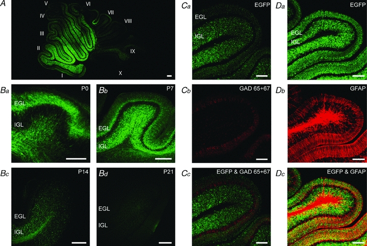
Figure 1. The 5-HT3 receptor is transiently expressed by glutamatergic granule cells in the cerebellum.

A, EGFP expression pattern in a P8 5-HT3A/EGFP transgenic mouse. Relatively high expression can be found in lobules I–VI. Roman numerals indicate cerebellar lobules. B, EGFP expression pattern in 5-HT3A/EGFP transgenic mice at P0 (Ba), P7 (Bb), P14 (Bc), and P21 (Bd). The expression pattern follows the migration pathway of the cerebellar granule cells from the external (EGL) to the internal (IGL) granule cell layer, with diminished expression from P14 onwards and no expression after P21. C, immunostaining of GAD65 + 67 in EGFP-expressing cerebellum (Ca–Cc) from a P7 5-HT3A/EGFP transgenic mouse shows no coexpression of 5-HT3 receptors with GABAergic neurons. D, immunostaining of GFAP in EGFP-expressing cerebellum (Da–Dc) from a P7 5-HT3A/EGFP transgenic mouse shows no coexpression of 5-HT3 receptors with glial cells, further confirming the expression of 5-HT3 receptors by glutamatergic granule cells in the cerebellum. Scale bars indicate 100 μm.
