
Figure 2. The reduction in bAP-evoked [Ca2+]i increase is not due to a change in the AP peak amplitude or width at the site of the measurement.
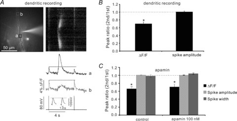
A, the image shows a dendrite with a patch electrode about 70 μm from the soma. The uncaging pulse at that location caused a reduction in the bAP Ca2+ signal at the site of the evoked Ca2+ wave (continuous ROI, a) but not at a distal location (dotted ROI, b) where the wave did not reach. The inset in the electrical trace shows that there was no change in the AP peak amplitude or shape. The cell was loaded with 50 μm OGB-1. B, summary histogram (*P < 0.01; two-tailed t test; n = 4). C, summary histogram for experiments showing that 100 nm apamin did not affect synaptically evoked Ca2+ wave-mediated suppression or spike parameters (*P < 0.01 for control; *P < 0.01 for apamin; two-tailed t test; n = 4). Ca2+ measurements from dendrites; electrical measurements from soma.