
Figure 5. Synaptically evoked NMDA spikes suppress bAP-evoked [Ca2+]i increases on the oblique dendrites.
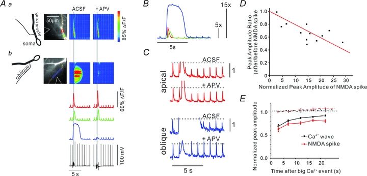
A, an experiment similar to that in Fig. 1 except that the low-affinity indicator OGB-5N (200 μm) was used. The top part shows ROIs and pseudocolour images of bAP signals and a synaptically evoked (100 Hz for 500 ms, grey vertical bar) Ca2+ wave along the main apical dendrite. The lower images show the same experiment except a rectangular ROI and pixel line along an oblique dendrite were selected. The tetanus evoked a larger and slower [Ca2+]i increase at these locations. Repeating the experiment in the presence of 100 μm APV blocked the large Ca2+ signal on the oblique dendrites but had no effect on the Ca2+ wave on the apical shaft. B, fluorescence signals at the three ROIs normalized to the amplitude of a bAP signal generated before the tetanus. The wave was about 5× the bAP signal and the NMDA spike more than 15× the bAP signal. C, high-resolution measurements showing that the Ca2+ wave suppressed the bAP signal on the apical shaft and the NMDA spike suppressed the signal on the oblique dendrite. In the presence of APV the suppression on the oblique dendrite did not occur. D, the suppression amplitude was plotted against the [Ca2+]i increase from the NMDA spike at a specific location, normalized to the amplitude of the bAP signal evoked at that location before the NMDA spike was generated. Larger reductions were measured at locations with the largest normalized NMDA spike-generated [Ca2+]i increases. The red line is the best fit to the data, with the line forced to 1.0 when the [Ca2+]i change was 0. Only experiments using 200 μm OGB-5N are included in the plot. E, summary (n = 5, NMDA spike; n = 5, Ca2+ wave). The time window for the suppression was similar on the oblique dendrites (NMDA spike) and on the apical shaft (Ca2+ wave). The amplitude of the bAP-evoked transients did not recover to initial values by the end of the 20 s trial. The dotted lines show the amplitudes of a series of bAPs evoked transients at 2 s intervals (normalized to the 1st transient) when there was no Ca2+ wave or NMDA spike. In both the apical dendrite (n = 5) and the oblique dendrite (n = 5) there was no change during the 20 s trial, showing that neither indicator bleaching, photodynamic damage nor change in bAP parameters had an effect on the transients during the trial period.