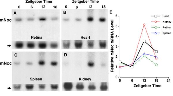
Figure 3.
mNoc mRNA is expressed rhythmically in C3H/He mouse retina (A), heart (B), spleen (C), and kidney (D) in a light dark cycle (LD). Tissues for RNA extraction were collected at Zeitgeber Times (ZT) 0 (24), 6, 12 and 18 with lights on at ZT 0 and off at ZT12. A through D are typical blots of mNoc for each tissue, and the lower panel is a hybridization of the same membrane with a β-actin probe. These blots are representative of three replicate experiments. In E phosphor imaging was used for quantitation of changes in mNoc mRNA level seen in A-D, standardized to β-actin. The minimum for each plot is one and the Y-axis shows the fold change.

