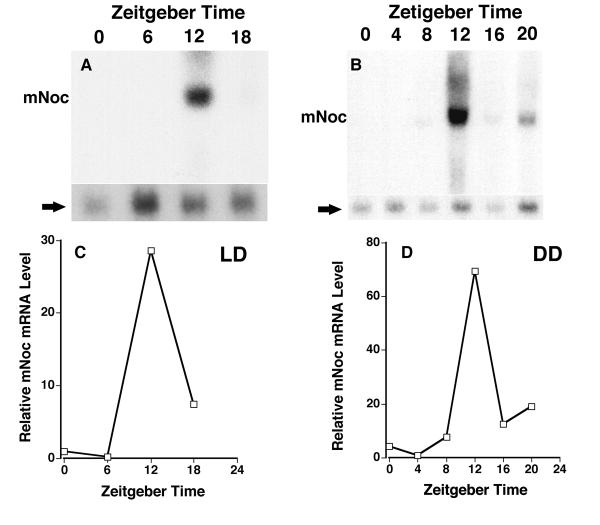
Figure 4.
mNoc mRNA in liver exhibits a high amplitude rhythm with peak expression at ZT12 in both LD (A and C) and DD (B and D). In LD samples were taken at 6 hour intervals as in Figure 4. Samples in DD were taken at Zeitgeber Times (ZT) 0 (24), 4, 8, 12, 16 and 20 referenced to the LD cycle immediately before DD treatment. Mice were in DD for 36 hours before beginning collections. The rhythmic changes illustrated are representative of three replicates for LD and two for DD. Phosphor imaging was used as in Figure 4E to quantitate mNoc mRNA levels (C and D). Note that the amplitudes of the rhythms are much higher in liver than for other tissues.

