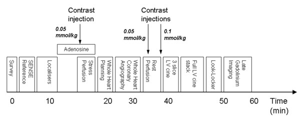
Figure 2.
CE-MARC cardiac magnetic resonance protocol. The protocol commences with a low-resolution survey scan and localisers. Intravenous adenosine is then administered for approximately 4 minutes at 140 mcg/kg/min, following which first pass stress perfusion imaging is undertaken after the injection of 0.05 mmol/kg dimeglumine gadopentetate. Three dimensional whole heart MR coronary angiography follows the low resolution coronary survey and free-breathing 4 chamber cine (used to assess slice coverage and diastolic coronary rest period respectively). Rest perfusion imaging is undertaken a minimum of 15 minutes following stress perfusion, with a further injection of 0.05 mmol/kg dimeglumine gadopentetate. A final injection of 0.1 mmol/kg dimeglumine gadopentetate is given following this sequence, bringing the overall gadolinium dose to 0.2 mmol/kg. Resting left ventricular function is then assessed, initially for three slices planned identically to the perfusion slices, and then for the entire left ventricle using contiguous slices. A modified Look-Locker inversion time scout is performed prior to late gadolinium enhancement imaging in short axis, vertical long axis and horizontal long axis orientations. Times indicated on the diagram are approximate and sequence blocks are not drawn to scale.

