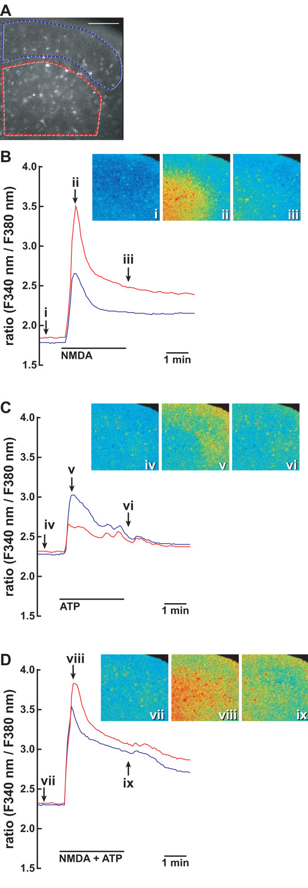
Figure 10.
Enhancement of NMDA-evoked [Ca2+]i increase by ATP in the spinal cord. A. Fluorescence image of a transverse section of fura-2-loaded L5 spinal cord excited at 340 nm. Areas surrounded by blue and red lines depict the regions to monitor fluorescence images of the dorsal horn. B-D represent typical traces of two regions showing the enhancement and prolongation of NMDA-evoked [Ca2+]i increase by ATP. The slice was serially stimulated by 100 μM NMDA (B), 100 μM ATP (C), and 100 μM NMDA and 100 μM ATP (D). Between stimulation, the spinal slice was perfused by the Krebs solution for more than 30 min. [Ca2+]i changes in spinal slices were fluorometrically monitored at 5-s intervals. [Ca2+]i changes are expressed as a ratio of fluorescence emission intensity excited at 340/380 nm. Fluorescence images (i)-(ix) are shown in pseudocolour as ratio images at the indicated point on [Ca2+]i traces (B-D). Similar results were obtained with 10 slices prepared from 5 mice. Bar = 100 μm.

