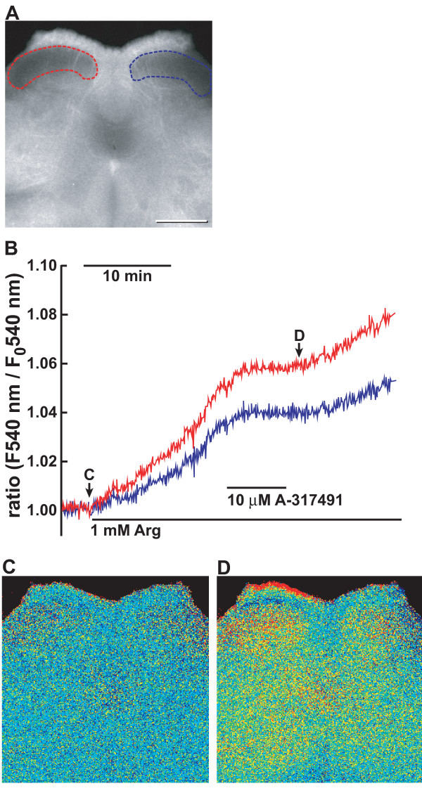
Figure 11.
Effect of A-317491 on NO production in the spinal cord 7 days after L5 spinal nerve transection. A. Fluorescence image of a transverse section of DRA-4M-loaded L5 spinal cord excited at 540 nm. Areas surrounded by red and blue lines depict the regions to monitor fluorescence of the dorsal horn in the sides ipsilateral and contralateral to L5 spinal nerve transection. B-D. Inhibition of NO formation by A-317491 in the dorsal horn of neuropathic pain model mice. Spinal slices were prepared from the L5 segment of neuropathic pain model mice 7 days after L5 spinal nerve transection. Fluorescence imaging for NO was obtained in the spinal slice loaded with DAR-4M and images were taken at 5-s intervals as described in "Methods". B shows representative traces of inhibition NO formation by A-317491 in the spinal slice prepared from neuropathic pain model mice. Underlines indicate the presence of 10 μM A-317491 and 1 mM L-arginine in the Krebs buffer. C, D. Fluorescence images of DAR-4M-loaded spinal cord at (C, an arrow) and 25 min after (D) the replacement of 1 mM L-NAME with 1 mM L-arginine. Fluorescence images are shown in pseudocolour as ratio images on the basis of the initial intensity at the start of measurement. Similar results were obtained with 5 slices of 5 animals. Bar = 500 μm.

