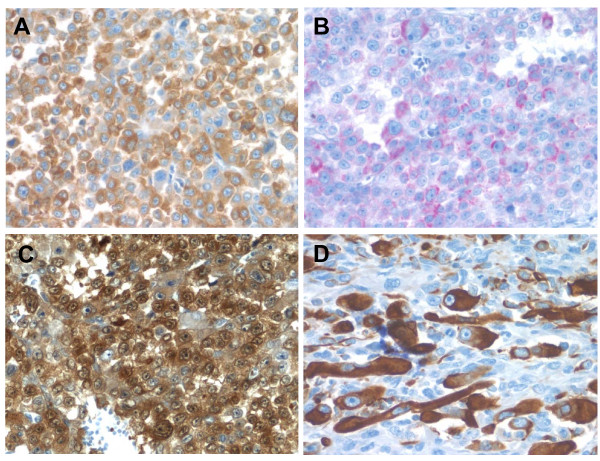
Figure 4.
Adrenocortical carcinosarcoma immunohistochemistry. A. The carcinomatous areas are strongly positive for synaptophysin (40×). B. Melan-A shows patchy positivity in the carcinomatous areas (40×). C. Calretinin immunostain showing diffuse cytoplasmic and nuclear positivity in carcinomatous areas (40×). D. Desmin immunostaining highlighting rhabomyoblastic cells in sarcomatous area (40×).

