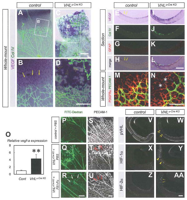
Fig. 4. Vascular defects in VHLα-Cre KO mice are attributable to ectopic VEGF expression.
(A–L) Whole-mount (A–D) or section (E–L) in situ hybridization for VEGF combined with immunostaining with indicated antibodies. Although VEGF expression is detected in astrocytes located in avascular area (arrows) of control mice, abundant VEGF expression is detected in the deep retinal layer (open arrowheads) where persistent hyaloid vessels invaginate in VHLα-Cre KO mice. (M,N) Immunostaining with PEC AM-1 (green) and PDGFRα (red) on P6 retinas. Despite normal astrocyte plexus (asterisks), vessel regression (open arrowheads) occurs in VHLα-Cre KO mice. (O) Quantitative PCR of vegfa for isolated RNA from P6 retinas (n = 6). (P–U) Fluorescent microscopic images in P6 retinas perfused with FITC-dextran (P–R), and confocal images labeled with PECAM-1 (S–U). Flt1-Fc injection into the eyes of VHLα-Cre KO mice reduces collateral flow (arrows in Q) and vascular structures (arrows in U) that exist abundantly in VHLα-Cre KO mice injected with vehicle (open arrowheads in Q,T). (V-AA) Immunostaining with indicated antibodies for sections of P6 retinas. While pVHL-expression is greatly reduced (open arrowheads in W), HIF-1α-immunoreactivity is increased (closed arrowheads in Y) in the deep retinal layer of VHLα-Cre KO mice. HIF-2α staining is detected in invaginating hyaloid vessels in VHLα-Cre KO (arrows in AA) Scale bars: 500 μm in (A–L). 200 μm in (P-AA); 100 μm in (M,N). **P < 0.01. All error bars indicate mean ± s.d.

