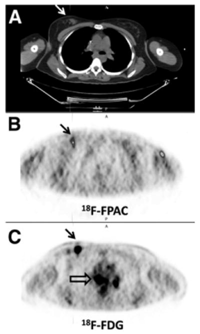
FIGURE 6.

Breast cancer patient with biopsy-proven tumor in right breast mass (arrows on CT [A] and PET [B and C] scans) and biopsy-proven sarcoid in mediastinum or hila. Increased 18F-fluoropacli-taxel (B; SUV uptake corresponding to tumor and no 18F-fluoropaclitaxel uptake corresponding to mediastinum/hilar lesions) seen on 18F-FDG scan (C; open arrow). 18F-FDG and 18F-fluoropaclitaxel images are scaled to SUVmax of 2.0. Tumor SUVmax on18F-fluoropa-clitaxel (B) at 78 min was 0.9; 18F-FDG (C) SUVmax at 123 min was 10.0. Uptake in anterior portion of left arm on 18F-fluoropaclitaxel image (B) is residual tracer within vessel wall. FPAC 5 fluoropaclitaxel.
