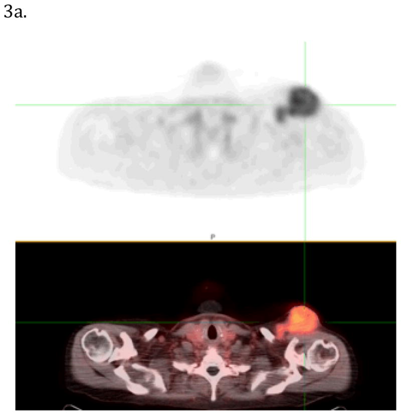
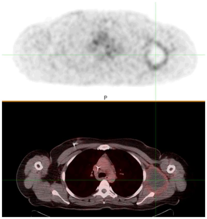
FIGURE 3.


Figure 3a and 3b. Images of α4vβ3 integrin PET/CT imaging performed after the injection of 18F-Flucilatide in two patients with melanoma. In FIGURE 3a., diffuse high uptake (SUVmax 6.4) in the large soft tissue metastasis can be seen (cross hairs). The metastatic melanoma focus in Figure 3b. shows a central photon deficit and mild (SUVmax of 3.0) 18F-Flucilatide uptake around the periphery
