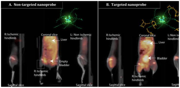
FIGURE 5.
Non-invasive PET/CT images of angiogenesis induced by hindlimb ischemia in a murine model. (A) Non-targeted dendritic nanoprobes. (B) Uptake of αvβ3 integrin targeted dendritic nanoprobes. Note accumulation of αvβ3 integrin targeted dendritic nanoprobes in the right ischemic hindlimb (box in B), when compared with the lack of uptake of the non-targeted agent (box in A). The structure and biodegradably nature of this nanoprobe may make it useful in developing a non-toxic, targeted combined therapy/imaging agent.
Reproduced with modifications, from Almutairi A, Rossin R, Shokeen M, et al. Biodegradable dendritic positron-emitting nanoprobes for the noninvasive imaging of angiogenesis. Proc Natl Acad Sci U S A. Jan 20 2009;106(3):685–690. with permission.

