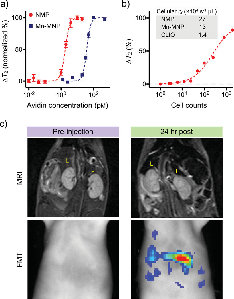
Figure 4.
Biological utility of NMPs. a) Small molecule detection was demonstrated using the avidin–biotin system as a model. Biotinylated NMPs were mixed with varying amounts of avidin, which caused aggregation of NMPs and corresponding changes in T2 relaxation times. The high r2 relaxivity of NMPs enabled extremely sensitive detection (≈1 pm of avidin), whereas the detection sensitivity with Mn-MNPs was ≈20-fold lower (20 pm). b) Cancer cells (SkBr3) targeted with HER2/neu-specific NMPs could be detected in 1 µL sample volumes, and the detection limit was near single-cell level. The inset table compares the cellular relaxivities of different particle preparations. c) NMPs incorporating near-infrared dyes (NMP-VT680) were used as dual in vivo imaging agents. Mice received intravenous injections of NMP-VT680 before undergoing both magnetic resonance imaging (MRI) and fluorescent-mediated tomography FMT). Due to large amounts of phagocytic cells, the liver (L) showed decreased signal intensity with MRI, while under FMT, the liver showed high fluorescent signals.

