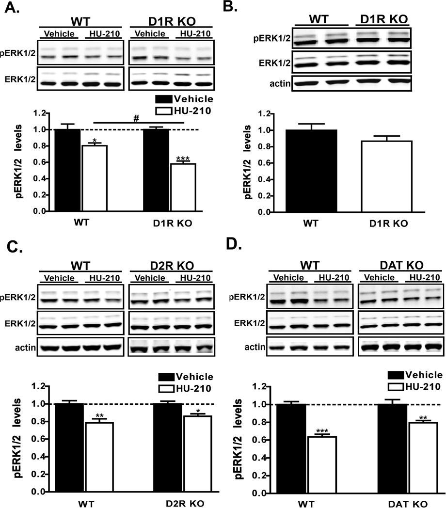
Figure 4. HU-210-mediated ERK1/2 inactivation is augmented in D1R KO mice.
A) Western analyses of pERK1/2 levels in extracts from dorsal striatum of wild-type (WT) or D1R KO mice that were administered HU-210 (0.25 mg/kg i.p.) and euthanized 60 min post-injection. n = 9–10 mice for each treatment group. B) Measurement of basal pERK1/2 levels between WT and D1R KO drug-naïve mice. pERK1/2 levels were normalized to the levels of ERK1/2 in dorsal striatal extracts. Data are mean ± S.E.M.; n = 5 mice for each group. An unpaired t-test found no significant difference between genotypes. C–D) HU-210 (0.25 mg/kg i.p.) was administered to WT, D2R KO (C) or DAT KO (D) mice and the levels of pERK1/2 in striatal extracts was measured by western blot 60 min post-injection. Data are mean ± S.E.M.; n = 4–5 mice for each treatment group. ***p < 0.001 and **p < 0.01 and *p < 0.05, vehicle versus HU-210 treatment within genotypes and #p < 0.05 for comparison between genotypes of the drug effect by two-way ANOVA.

