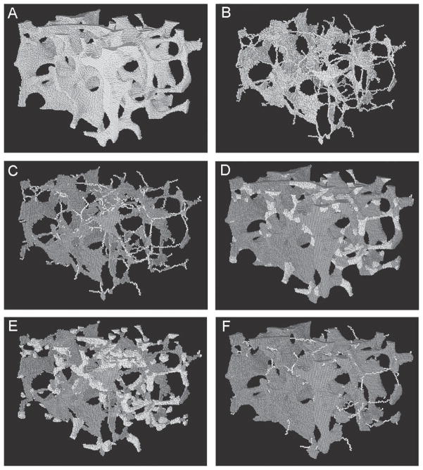
FIG. 1.
Results of DTA and reconstruction procedure on image of vertebral trabecular bone sample (3.2 × 3.2 × 2.1 mm3). (A) An original μCT image (full voxel model) of a trabecular bone sample. (B) Skeletonized image (skeleton model) of A. (C) Results of topological classification of B. Plate voxels are shown as dark gray, rod voxels in lighter shading. (D) Reconstructed structures with the trabecular type labeled for each voxel. (E) Rod-reconstructed model. (F) Plate-reconstructed model.

