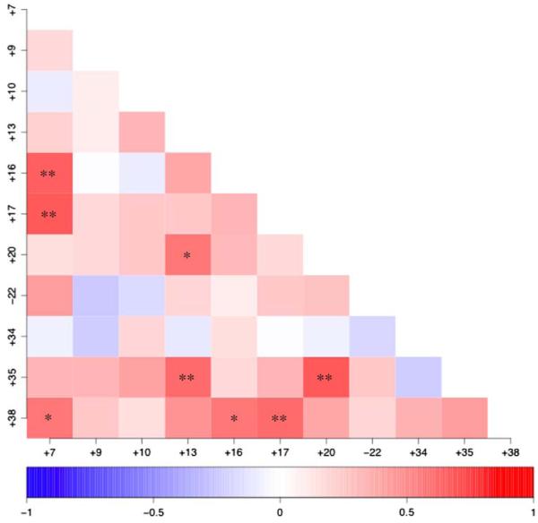
Fig. 3.
Correlation analysis of recurrent chromosome aberration in gliomas. Cases were scored according to the presence or absence of each pairwise combination of the 11 chromosome aberrations that were observed in ≥40% of the glioma population. The degree of correlation between aberrations is indicated on a scale of red (positive correlation) ↔ blue (negative correlation). Asterisks indicate pairwise combinations of chromosome aberrations that show significantly strong association i.e. they occur at a frequency that is significantly different from that predicted on the basis of their individual frequency in the population (* indicates p < 0.02, ** indicates p < 0.01)

