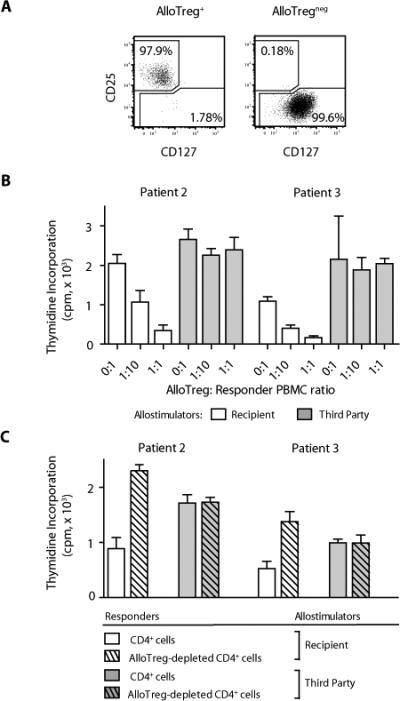
Fig. 2.

CD4+ Treg cells from recipients of alloanergized transplants suppressed recipient-specific alloresponses. (A) Flow cytometric sorting of patient PBMCs after transplant yielded ~98% pure populations of CD4+CD25hiCD127lo Treg cells (left) and Treg-depleted CD4+ cells with minimal (<0.2%) residual Treg cell content (right). Dot plots are gated on viable CD3+CD4+ cells. (B) Effect of addition of purified Treg cells from patient peripheral blood after transplant on mean (± SD) proliferation (thymidine incorporation) of fresh donor PBMCs stimulated with recipient (patient) or third-party allostimulators. Results are shown for patient 2 (day 43) and patient 3 (day 42). cpm, counts per minute. (C) Mean proliferation (± SD) of Treg-replete and Treg-depleted CD4+ cells from peripheral blood of patients 2 and 3 after transplant stimulated with recipient or third- party allostimulators. Results are shown for patient 2 (day 43) and patient 3 (day 42).
