
Fig. 2. Southern blot analysis of the fadD5 mutant strain and growth kinetics in standard medium.
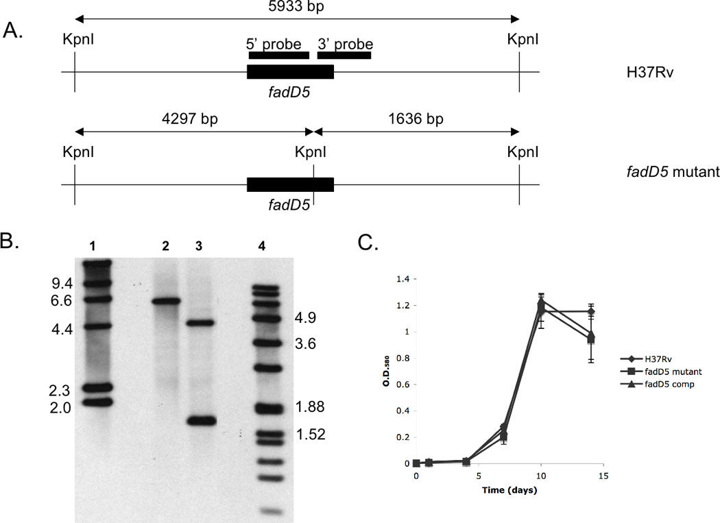
(A) Restriction digestion sites within and surrounding fadD5 genomic region. The 5’ and 3’ probes (black boxes) represent the regions recognized in the Southern blot analysis of H37Rv WT (top) and fadD5 mutant (bottom) genomic DNA. (B) Southern blot analysis of the WT (lane 2) and fadD5 mutant genomic DNA (lane 3) cleaved by the restriction enzyme, KpnI. The lanes are flanked by the digoxigenin-labeled molecular mass standards, II (lane 1) and VII (lane 4) (Roche Diagnostics). (C) Growth kinetics of the WT, fadD5 mutant, and fadD5 complemented strains in 7H9 medium. Cultures were inoculated in triplicate at O.D.580 0.004 and optical densities were measured at 1,4,7,10, and 14 days post-infection.