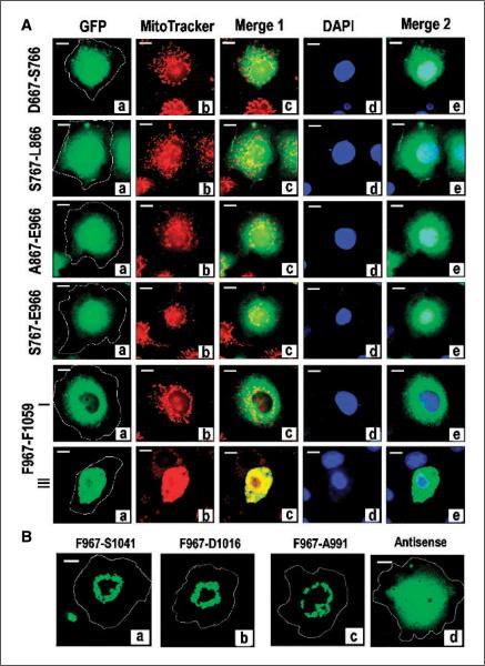
Figure 5.
Identification of an independent MAGD domain. A, analysis of GFP-tagged constructs coding for subdomains of C19ORF5C. COS cells were transfected with the indicated constructs with GFP at the NH2 terminus and cells were stained with MitoTracker and DAPI dyes. Cultures were examined for type I to IV phenotypes 24 hours after transfection in the same way as described for GFP-C19ORF5C. Yellow in Merge 1 and cyan in Merge 2 indicate overlap of green GFP-C19ORF5C with mitochondria (red) or DNA (blue), respectively. Representative cell from each transfected culture. Type II to IV cell morphologies were only observed in cultures transfected with F967-F1059 at the relative frequencies described for GFP-C19ORF5C in Fig. 1. Representative type I and III cell. B, reduction of the MAGD domain to a 25-residue sequence. The indicated constructs of MAGD domain-containing GFP-F967-F1059 with sequential deletions at the COOH terminus were tested for induction of the type II to IV phenotypes indicative of MAGD activity. Antisense, coding sequence for GFP fused to the in-frame antisense coding sequence for F967-A991.

