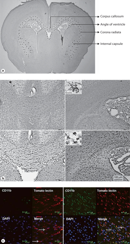
Fig. 3.
a Schematic representation of the distribution of activated microglia in the newborn rabbit following intrauterine endotoxin administration. The image represents a coronal section of a newborn rabbit brain under low magnification. The distribution of activated microglia in the endotoxin-treated kits is indicated schematically by black dots in the corpus callosum, corona radiata and angle of the ventricle. At this level, microglia are found along the border of the ventricles, and in the corpus callosum, corona radiata and internal capsule. b Microglial staining in neonatal rabbits on day 1 of life. Slides were stained for microglia using Lycopersicon esculentum (tomato lectin). An intense increase in numbers of microglia and a change in morphology of the microglia to a more rounded and amoeboid form is noted in the corpus callosum, around the lateral ventricles and in the corona radiata in endotoxin kits when compared with controls. Left: Corpus callosum. Right: Angle of ventricle. Upper row: Control. Lower row: Endotoxin. Scale bar = 100 μm. Insets High-magnification image of the microglia, demonstrating the ramified morphology in controls (upper inset) and a more rounded and amoeboid/bushy morphology in the endotoxin kits (lower inset). Scale bar = 10 μm. c CD11b expression in the neonatal rabbit brain on day 1 of life. Representative sections from control (left) and endotoxin kits (right). Increased expression of CD11b in the periventricular region of endotoxin kits is shown when compared with the control saline kits on day 1 of life. Microglia labeled with tomato lectin (red fluorescence) colocalized with CD11b (green fluorescence) predominantly in the endotoxin kits and demonstrated a more rounded morphology (arrows, right), while microglial cells in the control kits were mostly ramified (arrows, left) and did not stain for CD11b. Scale bars = 20 μm.

