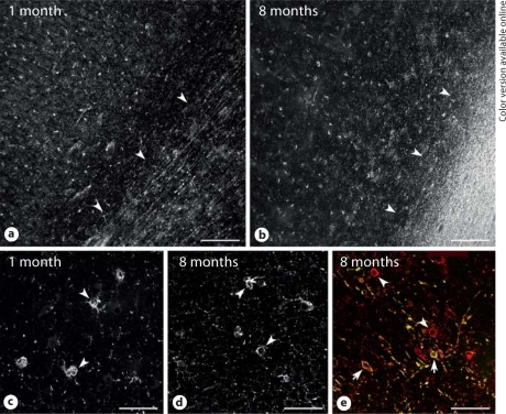
Fig. 6.
OL maturation is less pronounced in human parietal cortex compared with white matter. Low-power photomicrographs show numerous cells labeled with bO4 antibody at 1 month (a) and 8 months (b) after full-term birth. Note that the bO4-labeled cells in the cortex were preOL that did not stain for the O1 antibody (not shown). a, b The approximate boundary between cortical layer 6 and the subcortical white matter is indicated by the arrowheads and was confirmed by a Hoechst fluorescent counterstain (not shown). c, d Higher-power details of the preOL (arrowheads) from the cases in a (c) and b (d) demonstrate the simplified arbor of processes associated with the cortical preOL at both ages. e Both preOL (red, online version only; arrowheads) and immature OL (yellow, online version only; arrows) were visualized in a region of early myelination at the edge of the densely myelinated tract shown in b. Scale bars = 200 μm(a, b) and 50 μm(c–e).

