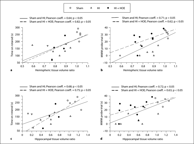
Fig. 2.
Correlation of ipsilateral hemispheric tissue volume and hippocampal tissue volume with motor and spatial learning in mice after HI. The accelerating rotarod test and MWM probe trial were performed at P30 and P60 after HI, respectively. Hemispheric and hippocampal tissue volume ratios of these animals were determined by T2-weighted MRI at P90. Correlations are presented for sham/HI and sham/HI + HOE animals. a Correlation of ipsilateral hemispheric tissue volume ratio with time spent on the rotarod. b Correlation of ipsilateral hemispheric tissue volume ratio with time spent in the training quadrant (MWM probe trial). c Correlation of ipsilateral hippocampal tissue volume ratio with time spent on the rotarod. d Correlation of ipsilateral hippocampal tissue volume ratio with time spent in the training quadrant (MWM probe trial).

