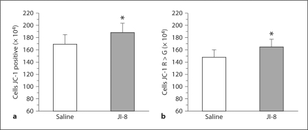
Fig. 5.
Flow cytometric assessment of mitochondrial function at 72 h using JC-1 in whole brain slices from HI rabbit fetuses treated with the nNOS inhibitor JI-8 compared with saline. * p < 0.05. a Significant increase in total number of cells stained with JC-1 in the JI-8-treated group compared with saline. b Significant increase in number of cells staining more red than green with JC-1 in slices treated with JI-8 compared with saline.

