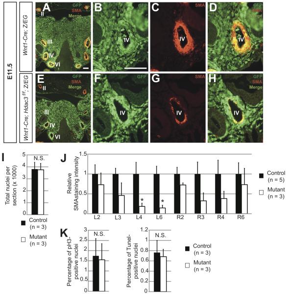
Figure 4. Hdac3Wnt1NCKO embryos display deficiencies in aortic arch artery smooth muscle at E11.5.
A-H. Immunohistochemistry for GFP and α-smooth muscle actin (SMA) in frontal sections of E11.5 pharyngeal arches. A, Representative image of a control pharyngeal arch at low magnification, showing robust layers of smooth muscle surrounding aortic arch arteries II, III, IV and VI (labeled), bilaterally. Note that the bulk of pharyngeal mesenchyme is comprised of GFP-positive neural crest derivatives. B-D, High magnification images of the left fourth arch artery in the control embryo. B, Neural crest cells populate the region directly surrounding the artery. C,D, A layer of neural crest-derived, SMA-positive smooth muscle surrounds the control artery. E, Low magnification image of a mutant pharyngeal arch, showing neural crest derivatives populating the mesenchyme. Note the decreased smooth muscle in the regions surrounding the third, fourth and sixth arteries, particularly on the left side, while a thick layer of non-neural crest-derived smooth muscle surrounds the second arteries, as in the control F-H, High magnification images of the mutant left fourth artery. F, Neural crest cells appropriately populate the region surrounding the artery. G,H, In affected mutants, few neural crest-derived cells surrounding the fourth arch artery are SMA-positive smooth muscle cells. Three of four mutant and zero of four controlembryos analyzed at this time point showed a similar pattern of staining. Roman numerals denote aortic arch artery number. I, Quantification of cells in the pharyngeal mesenchyme of aortic arches III-VI from serial sections of E11.5 control and mutant embryos. J, Quantification of SMA staining intensity of each aortic arch artery, from serial sections in control and mutant embryos. Asterisks denote p < 0.05. K, Quantification of phospho-histone H3 (pH3)- and Tunel-positive in serial sections of the pharyngeal arch mesenchyme. Scale bars: 100μm.

