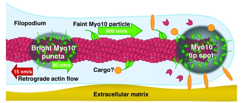
Fig. 3.
Myo10 undergoes several forms of intrafilopodial motility. Model of Myo10 movements in filopodia. In HeLa cells, bright puncta or clusters of Myo10 at tips of filopodia occasionally move rearwards within filopodia at the retrograde flow rate of ~15 nm/second. Bright puncta (areas of gray shading) have also been observed moving forwards within filopodia at ~80 nm/second. These puncta might include as-yet-undiscovered scaffolding components, actin polymerization machinery or cargos. Faint Myo10 particles (green), probably corresponding to dimers, move much faster (~600 nm/second) towards the filopodial tips. Forwards movements require Myo10 motor activity, whereas rearwards movements are hypothesized to be due to coupling to actin (red) undergoing retrograde flow. Myo10-interacting proteins (orange) include both cytoplasmic and membrane-associated proteins. See supplemental movie 5 of Kerber et al. for an animated model of Myo10's intrafilopodial motility (Kerber et al., 2009).

