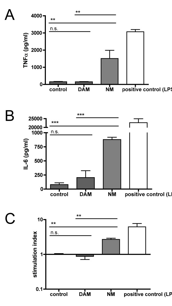
Figure 4.
NM-mediated dendritic cell (DC) activation is functional and independent of the melanin backbone. A, B: DCs were cultured for 48 h in the presence of medium alone, DAM, NM, or LPS (positive control). The amount of proinflammatory cytokines in the cell culture supernatants was determined by ELISA. A: TNF-α. B: IL-6. C: DCs treated for 48 h with medium alone, DAM, NM or LPS (positive control) were cocultured with allogenic T cells. A-C: Data from 2 independent experiments with triplicates and quadruplicates as mean ± S.E.M.; statistical analysis with one-way ANOVA using Bonferroni multiple comparison as post test; n.s. = non specific (p > 0.05), * p < 0.05, ** p < 0.01, *** p < 0.001).

