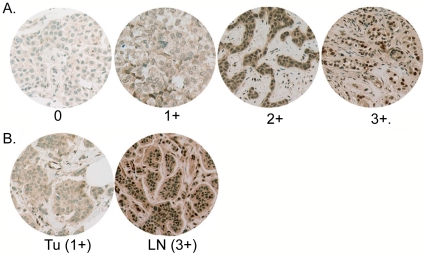
Figure 1. Tyrosine phosphorylated Stat3 in breast cancer and matched metastatic axillary lymph nodes.
A. Tissue microarrays (TMAs) of primary breast tumors (38) were analyzed for nuclear tyrosine-phosphorylated Stat3 (pStat3) by immunohistochemical (IHC) analysis. Examples of no detectable pStat3 (0), low levels (1+), moderate levels (2+) and high levels (3+) are shown. B. Comparison of pStat3 levels of tumor cells by IHC in matched primary tumors with axillary lymph nodes. 37% of primary tumors expressed 2–3+ pStat3, while 63% of matched lymph nodes expressed 2–3+ pStat3. Relative pStat3 IHC staining was greater in the lymph node compared to the corresponding primary tumor in 15/38 specimens, equal in 18/38 and less in 5/38. A representative example of a tumor (Tu) specimen with 1+ pStat3 versus 3+ pStat3 in the corresponding lymph node (Ln) is shown.

