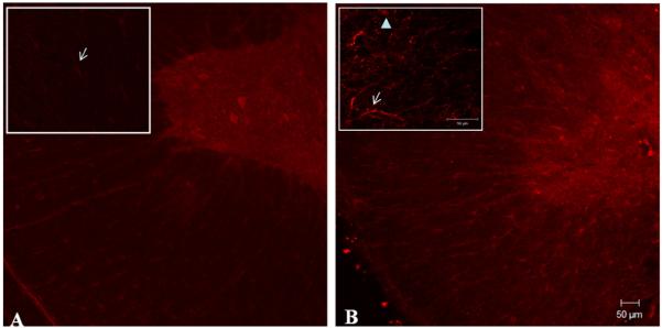
Fig. 2.
Immunohistochemical analysis of P2Y2 receptors in normal and contused spinal cord. Rabbit anti-P2Y2 receptor antibody was used in fixed adult spinal cord to determine the regions of protein expression in control (A) and injured (B) spinal cord samples (n = 3). Insert indicates the pattern of P2Y2 expression in regions of the white matter visualized at higher magnification. Arrows indicate astrocytes-like cells positively labeled in regions of the white and the arrow-head point out an axon-like structure (gamma = 0.7). Scale bar = 50 μm.

