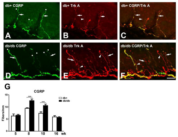
Fig. 3. Increased CGRP-positive IENFD in db/db mice during the period of mechanical allodynia.
A-C: Immunohistochemistry studies from the hind paw skin of 8 wk-old db/+ mice (A-C) demonstrated CGRP (A) and Trk A (B)-positive IENF. Each counted IENF was labeled with a white dot. A-C: CGRP immunoreactive fibers are also positive for Trk A (arrows). There are CGRP-positive but Trk A-negative IENF (B: arrowhead). C: The merged picture demonstrates both CGRP-positive (arrows) and -negative (arrowhead) Trk A-positive IENF. D-F: Immunohistochemistry studies from the hind paw skin of 8 wk-old db/db mice demonstrated SP (D) and Trk A (E)-positive IENF. D: CGRP-positive IENF (arrows) in epidermis of db/db mice. E: Trk A-positive IENF (arrow) in db/db mice. Some Trk A-positive IENF are negative for SP (arrowhead). F: The merged picture demonstrates both CGRP-positive (arrow) and -negative (arrowhead) Trk A-positive IENF. G: Quantification analysis of IENFD demonstrates increased CGRP-positive IENFD at 8 and 10 wk of age. ***, p < 0.001, N = 4. Bar = 20 μm.

