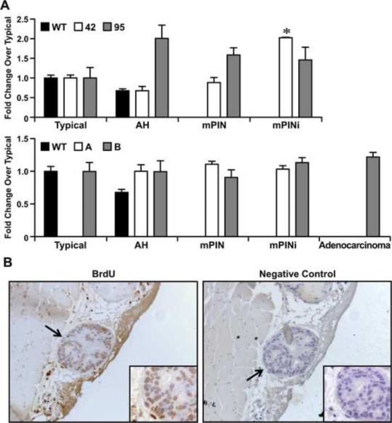
Figure 5. Ron overexpression in the prostate increases cell proliferation.
A, The extent of cell proliferation in prostates containing each pathology [Typical, atypical hyperplasia (AH), mPIN, mPINi, and adenocarcinoma] for the select transgenic line is depicted. The extent of proliferation observed in WT prostates with a typical pathology was normalized to 1 and the fold-change over typical pathology is depicted. Of note, as the severity of pathology increases, the extent of cell proliferation increases in the prostates of transgenic mice compared to wild type prostates. *P<0.05 compared to typical. B, Pictures showing representative staining of a transgenic prostate and a corresponding negative control. All four transgenic lines demonstrate increased proliferation at all histological stages when compared to wild type. Original magnification was taken at 40× and insets at 63×.

