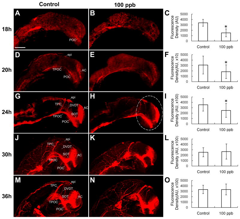
Figure 2. Effect of Pb on density of axon tracts of embryonic zebrafish brain in lateral view at 18, 20, 24, 30 and 36 hpf.
Anti-acetylated α-tubulin staining clearly showed axon tracts at 18, 20, 24, 30 and 36 hpf. A, B and C: 18 hpf (n=15–18); D, E and F: 20 hpf (n=14–16); G, H and I: 24 hpf (n=14–16), J, K and L: 30 hpf (n=21–22); M, N and O: 36 hpf (n=13–14). Density data of stained region of interest (white dash line marked) were measured using Image-pro plus (C, F, I, L and O). The units for F are 10 times greater than C while the units for I, L and O are 100 times greater (denoted as ×10 and ×100). AC, anterior commissure; POC, post-optic commissure; SOT, supra-optic tract; TPOC, tract of post-optic commissure; TPC, tract of the posterior commissure; DVDT, dorsal-ventral diencephalic tract; epi, epiphyseal cluster. Scale bar=100 μm. *p<0.05.

