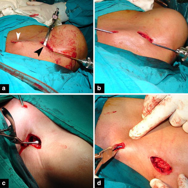
Fig. 2.

Operative photos of the RTEN technique. a A small incision is made over the fracture site (black arrowhead). The medial fragment is grasped with bone forceps and lifted up in the wound. The medullary canal is reamed with a drill bit with a diameter similar to the nail (usually a 2.5 mm bit is required). When the drill bit has penetrated the anterior cortex and is felt under the skin, a tiny incision is made over it. b A nail is inserted through the medullary canal and allowed to come out through the small medial incision. c The fracture is reduced and the nail is pushed inside the lateral fragment under fluoroscopic control. d Bending of the tip of the nail before wound closure
