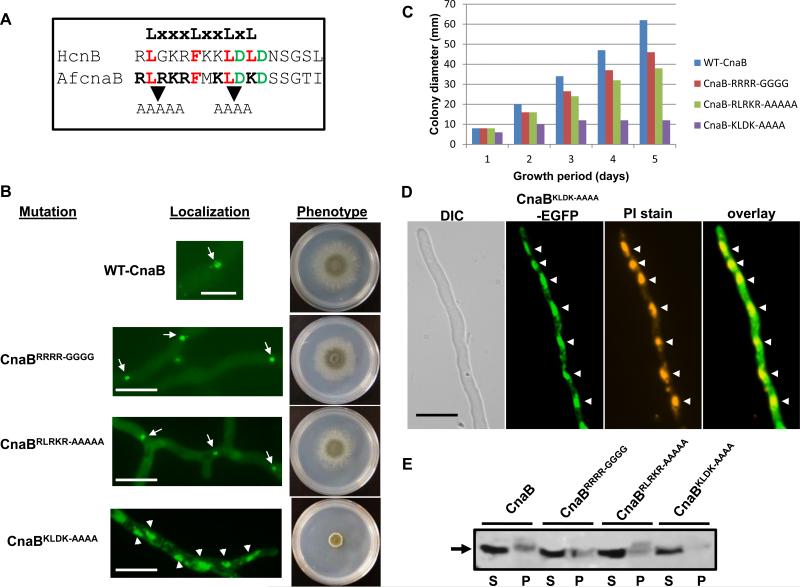
Figure 12.
(A). Alignment of the predicted nuclear exit signal sequence (NES) at the N-terminus of human CnB1 and A. fumigatus CnaB is shown along with the NES consensus motif. Conserved residues in the LxxxLxxLxL motif are shown in red and other favored acidic residues are in green. Mutations in the RLRKR and KLDK motifs are indicated (B). Expression of mutated constructs of N-terminal basic motifs (15-ARRRA-20 to 15-AGGGGA-20, 44-RLRKR-48 to 44-AAAAA-48 and 51-KLDK-54 to 51-AAAA-54) in A. fumigatus CnaB by tagging with egfp. Note that while the control WT-CnaB, CnaBRRRR-GGGG and CnaBRLRKR-AAAAA expressing strains showed localization at the septa (indicated by arrows), the CnaBKLDK-AAAA strain showed nuclear localization (indicated by arrowheads). Hyphal growth recovery in the strains was observed by inoculating a total of 1x10⁴ conidia GMM agar medium and incubating at 37°C for 3 days. WT-CnaB (blue bar), CnaB-SW2 (red bar), CnaB-tB3 (green bar). (C). Measurement of colony diameter of the strains expressing WT-CnaB (blue bar), CnaBRRRR-GGGG(red bar), CnaBRLRKR-AAAAA (green bar) and the CnaBKLDK-AAAA (purple bar) strains over a period of 5 days. For radial growth quantification the strains were grown on GMM agar medium in triplicate and values are depicted as average colony diameter. (D). Nuclear staining of the strain expressing CnaBKLDK-AAAA tagged to egfp. Localization of the CnaBKLDK-AAAA in the nucleus was confirmed by propidium iodide staining of the nuclei. Arrowheads indicate the nuclei. Scale bar 10 μm. (E). Detection of the expression of mutated CnaB-EGFP fusion proteins. Total proteins (~50 μg) extracted from the strains cultured for 24 h were subjected to 12.5% SDS-PAGE and Western analysis using anti-GFP rabbit polyclonal primary antibody and peroxidase labeled anti-rabbit IgG secondary antibody. Detection was performed using the SuperSignal West Pico chemiluminescent substrate. S and P indicate supernate and pellet fractions, respectively.

