
Figure 6. Role of IMAC in RIRR and arrhythmias in intact myocardium.
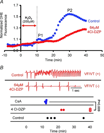
A, average normalized O2− responses of control (blue, n = 4) and 64 μm 4′-Cl-DZP (red, n = 4)-treated hearts to perfusion with 200 μm H2O2 for 10 min. In the treatment group, 4′-Cl-DZP perfusion was introduced 10 min prior to the H2O2 challenge and maintained throughout the entire protocol. The timing of H2O2 challenge is indicated by the red arrow. B, representative volume-conducted ECG traces from control and 4′-Cl-DZP-treated hearts challenged with an identical H2O2 perfusion protocol indicating the presence and absence of sustained VT in control and 4′-Cl-DZP-treated hearts, respectively. Time of onset of VT/VF in control (black), CsA- (blue), and 4′-Cl-DZP- (red) treated hearts is plotted. Open red circles indicate absence of arrhythmias at the end of the experimental protocol in 4′-Cl-DZP hearts. 100% of CsA- and 50% of 4′-Cl-DZP-treated hearts exhibited VT/VF. Time of onset was earlier in CsA-treated hearts.