
Figure 10. ECE-1-mediated resensitization of SP-induced Ca2+ transients in myenteric neurones.
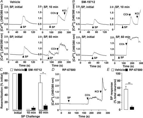
A–C, mouse myenteric neurones in culture were challenged with SP (initial, 10 nm, 5 min), washed, and the same neurones were re-challenged (10 nm SP) after 10 or 60 min recovery. After the final SP challenge, neurones were exposed to carbachol (CCh, 1 μm) and then KCl (50 mm). A and B, Ca2+ transients from the same neurones in each experiment (upper panel: 10 min re-challenge; lower panel: 60 min re-challenge) treated with vehicle (A) or SM-19712 (B). C, resensitization of neurones treated with vehicle or SM-19712. The proportion of neurones responsive to initial SP were recorded and are represented as 100% when compared to the proportion of the same neurones responsive to SP re-challenge. *P < 0.05 to vehicle, 46–102 neurones per group, n > 4 experiments (i.e. neuronal cultures from 3–5 mice per preparation). D and E, mouse myenteric neurones in culture were incubated with vehicle or the NK1R antagonist RP-67580, which suppressed detectable changes in [Ca2+]i. **P < 0.005 compared to vehicle, 149–176 neurones per group, n > 4 mice.